Cornea Clinic
Shankar’s eye hospitals Servicing best corneal department in Kollam District
we have well furnished super speciality cornea clinic and surgical unit that offers comprehensive and specialized care for corneal and Anterior eye diseases. Our team of expert corneal surgeons ,trained technical staff and skilled non technical staff handling therapeutic devices even the most challenging cases successfully.
The cornea is the clear, dome-shaped front surface of the eye. It plays a vital role in focusing light and enabling clear vision. However, the cornea can be affected by various conditions that can impair your sight and cause discomfort.
Super specialties Cornea Department
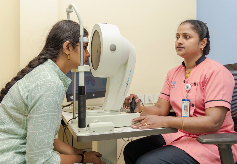
Diagnostic Equipment
- Shankar’s Eye Hospital invests in advanced diagnostic equipment specifically tailored for corneal evaluations.
- Most advanced Pentacam for evaluation of overall cornea within seconds, (we are offering German made Occulus Pentacam )
- Carl Zeiss Optic Coherence Tomography for evaluating Anterior part
- Japan made Topcon Speculomicroscopy using to check central corneal thickness
- The hospital’s commitment to providing comprehensive corneal services sets it apart, making it a beacon of eye care in the area.
Treatment for Corneal Diseases
Pterygium
Pterygium is a growth of reddish fleshy tissue on the cornea, usually forming on the nasal side and growing towards the pupilary area. It is twice as common in males as in females.
Symptoms of Pterygium
- May be asymptomatic
- Irritation
- Redness
- Foreign Body Sensation
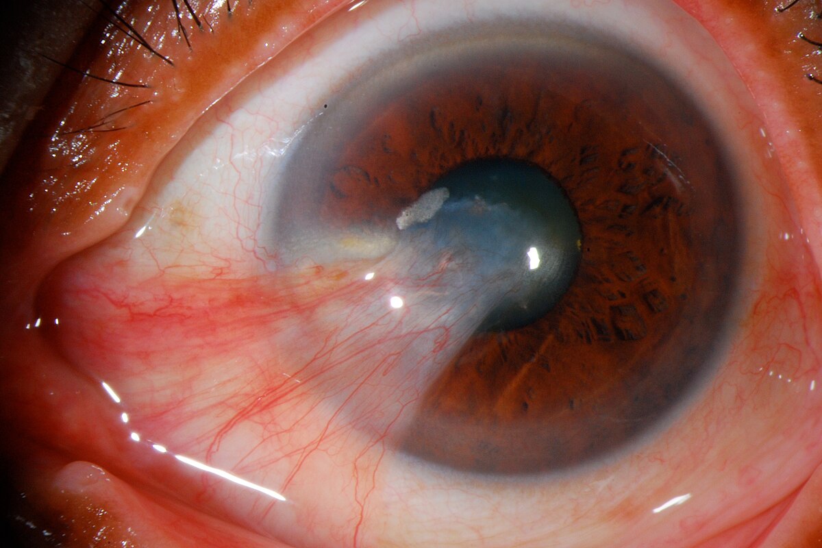
In extreme cases, it can cover your pupilary area and cause visual problems.
Risk Factors
- Sunlight exposure
- Hot & dry climate
- Age-related degeneration
- Hereditary
Prevention
You can prevent the development of a Pterygium by wearing sunglasses or a hat to shield your eyes from sunlight, wind, and dust. Using lubricant eye drops helps to prevent the growth of your Pterygium.
Treatment
If the Pterygium growth is large enough to cause discomfort or visual impairment, the final advice given by the surgeon is Pterygium removal. The surgery typically takes 15 to 20 minutes depending on the type of surgery:
- Simple excision
- Simple excision with auto conjunctival graft
- Simple excision with artificial amniotic membrane graft
- Glue
Keratoconus
A progressive disorder that causes the cornea to thin and bulge into a cone-like shape. This results in:
- Blurred vision
- Double vision
- Near-sightedness
- Astigmatism
- Sensitivity to light
Keratoconus usually affects both eyes and often develops in adolescence or early adulthood. About 7% of people with keratoconus have a family history of the condition. Some possible risk factors include eye rubbing and allergic eye diseases.
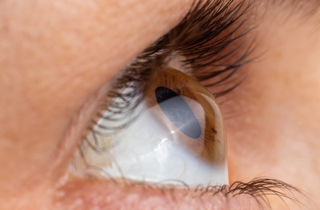
Shankar’s eye hospitals diligence a super specialityKeratoconus Clinic providing advanced diagnosis and management using modern instruments such as Pentcam, Specular Microscope, Anterior Segment Optical Coherence Tomography, etc.
TREATMENTS
- Spectacles for the mild cases
- Rigid gas permeable contact lenses
- C3R
- Scleral contact lenses
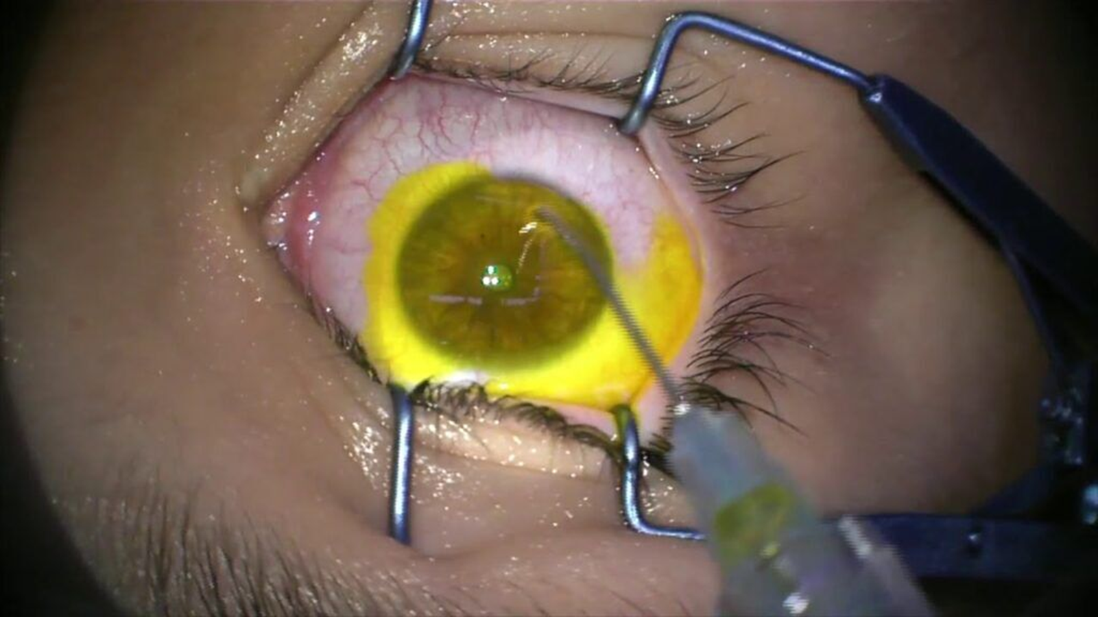
Shankar’s eye hospitals offering advanced surgery method for Keratoconus is Corneal Collagen Cross linking with Riboflavin
Corneal Ulcers
A corneal ulcer is a wound-like sore on your cornea. They are a medical emergency and need immediate care to prevent permanent eye damage, low vision, and blindness.
Infectious Causes
- Bacteria
- Viruses
- Fungi
- Parasites
Non-Infectious Causes
- Eye injuries
- Exposure
- Very dry eyes
- Toxic effects
- Immune conditions

- Post-Traumatic Treatment
- Stevens-Johnson Syndrome
- SEVERE DRY EYE
- Sjogren Syndrome
- Foreign Body (Superficial and Deep)

Investigations and Diagnostic Services
- Slit Lamp Bio microscopy
- Pentacam
- Stain test with fluorescein
- Swab test
We offer well equipped laboratory for swab and culture test to find out types of corneal ulcer.
As per the swab test result Ophthalmologists taking treatment as an emergency mode.
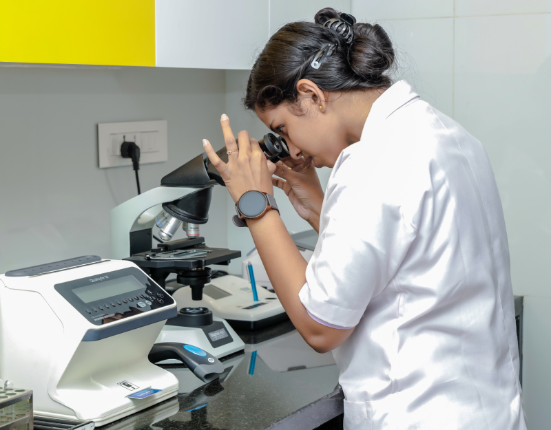
Infrastructure
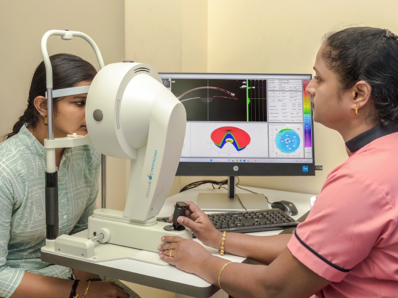
Pentacam
Provides a detailed 3D analysis of the anterior segment of the eye, crucial for diagnosing and managing corneal diseases.
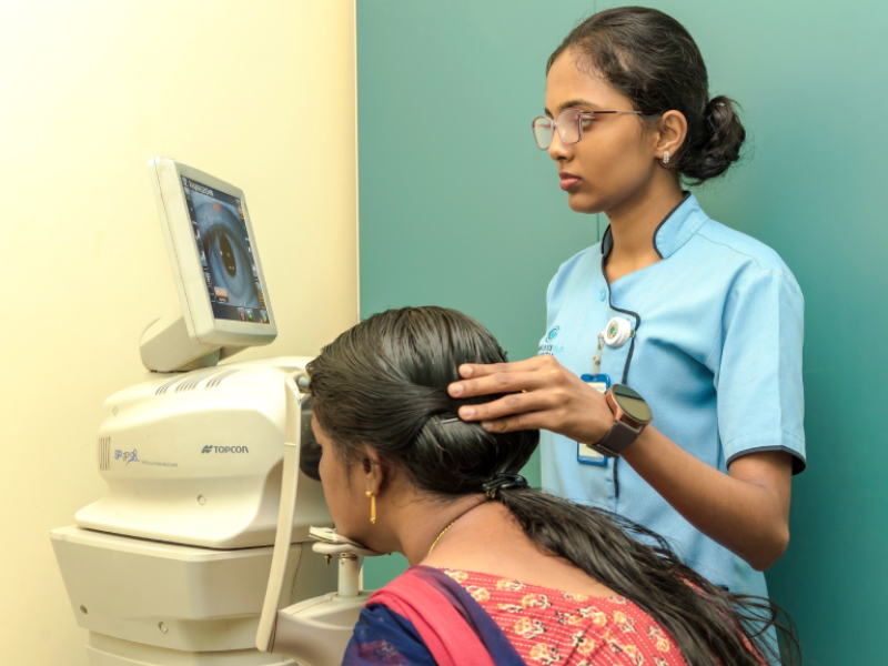
Specular Microscope
Evaluates the corneal endothelium to assess corneal health and detect early signs of disease.
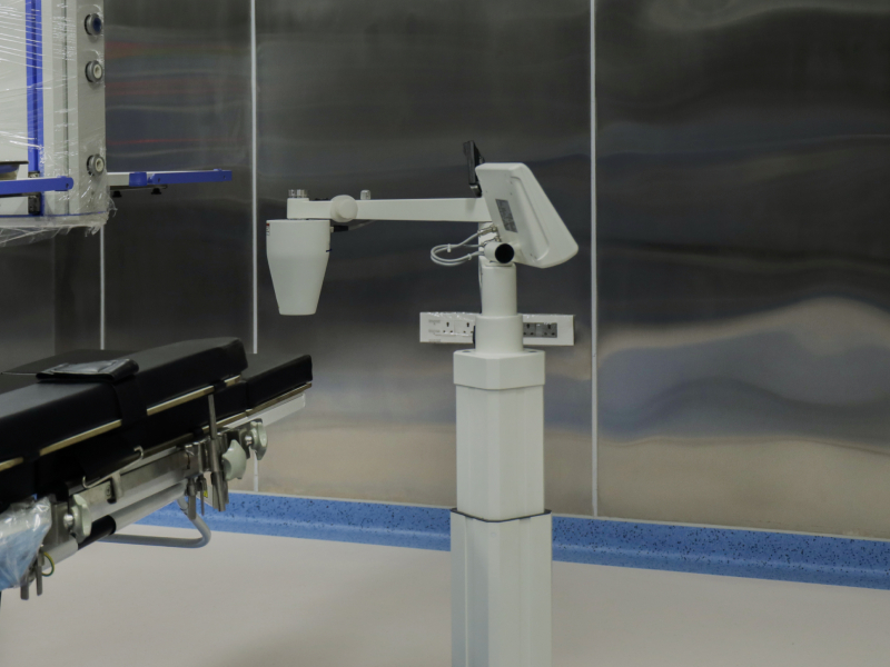
C3R- Corneal Collagen Cross-linking
It is a minimal invasive procedure used to prevent progression of corneal ectasia.In corneal cross-linking, doctors use eyedrop medication and ultraviolet light from a special machine to make the tissues in cornea stronger.C3r is the only treatment that can stop progressive keratoconus from getting worse